Mosaicing Tools
Open-source software tools to build mosaics of partially overlapping images


Mosaicing Tools
Open-source software tools to build mosaics of partially overlapping images

F. Piccinini, M. Pierini, E. Lucarelli, A. Bevilacqua, Semi-quantitative monitoring of confluence of adherent mesenchymal stromal cells on calcium-phosphate granules by using widefield microscopy images. Journal of Materials Science: Materials in Medicine, May 2014. Download
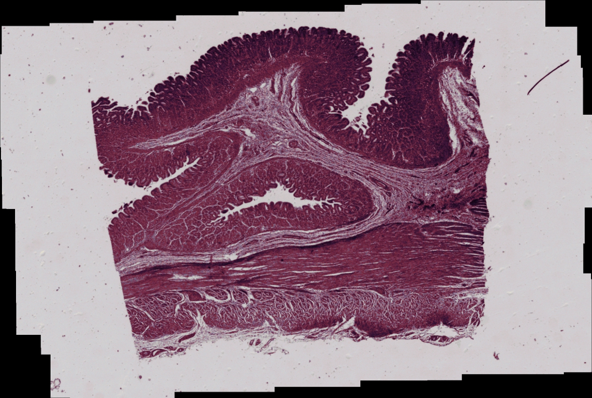
F. Piccinini, A. Bevilacqua, E. Lucarelli, Automated image mosaics by non-automated light microscopes: the MicroMos software tool. Journal of Microscopy, September 2013. Download
F. Piccinini, E. Lucarelli, A. Gherardi, A. Bevilacqua, Multi-image based method to correct vignetting effect in light microscopy images. Journal of Microscopy, October 2012. Download
F. Piccinini, A. Bevilacqua. Colour vignetting correction for microscopy image mosaics used for quantitative analyses. BioMed Research International, June 2018. Download
MicroMos Version 3.0 (*), License. The first version endowed with a Graphical User Interface (GUI) and a module for manually defining the shifts between the images to be stritched.
MicroMos Version 2.0 (*), License. To build mosaics of brightfield, phase-contrast and fluorescent images.
MicroMos Version 1.0 (*), License. To build mosaics of brightfield and phase-contrast images only. (click on "Supplementary Material" section)
F. Piccinini, A. Tesei, G. Paganelli, W. Zoli, A. Bevilacqua, Improving reliability of live/dead cell counting through automated image mosaicing. Computer Methods and Programs in Biomedicine, December 2014. Download
Sample set 01: 11 tiff images, 1600x1200 pixels, RGB, acquired in brightfield by using a 4x objective. Description: hemocytometer's grid with cells stained by using Trypan blue. The background of the images is particularly blue.
Sample set 02: 11 tiff images, 1600x1200 pixels, RGB, acquired in brightfield by using a 4x objective. Description: hemocytometer's grid with cells stained by using Trypan blue. The background of the images is really bright (over-exposed).
Sample set 03: 11 tiff images, 1600x1200 pixels, RGB, acquired in brightfield by using a 4x objective. Description: hemocytometer's grid completely empty (no cell seeded, no culture medium inside the grid).